 Breaking News & Live Updates
- Breaking News & Live Updates
- Breaking News & Live Updates
- Breaking News & Live Updates
- Breaking News & Live Updates
- Breaking News & Live Updates
- Breaking News & Live Updates
- Breaking News & Live Updates
- Breaking News & Live Updates
- Breaking News & Live Updates
AI Reveals Asymmetric Brain Aging and Alzheimer's Markers
A recent investigation published in the journal PNAS presents an innovative artificial intelligence technique for mapping the varied aging processes across different areas of the human brain. By conducting a detailed examination of MRI scans, the study offers compelling evidence that the brain's aging trajectory is not uniform, with particular regions displaying more pronounced biological aging in individuals afflicted with Alzheimer's disease.
Healthcare professionals and scientists frequently employ Magnetic Resonance Imaging (MRI), a method that uses potent magnetic fields to generate elaborate visualizations of internal bodily structures, to determine an individual's "brain age." This measurement indicates whether a person's brain appears biologically older or younger than their chronological age. A brain that exhibits characteristics of advanced age is often considered a risk factor for cognitive decline and various neurodegenerative conditions.
Uneven Brain Aging Across Hemispheres and Regions Uncovered by AI
Traditionally, researchers have relied on a singular, overarching brain age metric. However, the human brain is an intricate organ composed of distinct regions, each responsible for specialized functions. For instance, the frontal lobe is critical for complex decision-making, while the hippocampus plays a crucial role in memory formation. Given their differing roles, these areas tend to age at varying rates. A single brain age score can mask these regional disparities, making it more challenging to identify early indicators of localized damage. The study's lead author, Andrei Irimia, an associate professor of gerontology at the USC Leonard Davis School of Gerontology, explained that their previous work focused on global brain age, where AI estimated the brain's apparent age from an MRI. While useful as a summary, this approach condensed the entire brain's complexity into a single number, hindering biological interpretation. To address this, they developed a local brain age model, which not only assesses the overall brain's appearance but also meticulously evaluates the age of each individual part. This model transforms a structural MRI into a spatial map illustrating local brain age patterns.
This pioneering research, spearheaded by Nikhil N. Chaudhari alongside Irimia and their colleagues at the Irimia Lab, sought to construct a more granular map of brain aging. The team aimed to quantify biological aging at an exceptionally detailed level, dissecting the brain into minute three-dimensional units called voxels. This meticulous approach was intended to precisely pinpoint areas of accelerated brain aging and understand the relationship between these specific regional changes and cognitive decline. To develop their localized brain aging map, the scientists trained a deep learning computer model using MRI scans from 14,748 cognitively healthy adults ranging from 19 to 100 years old. Irimia emphasized the importance of scale and interpretability, noting that the model was trained on a vast and diverse dataset, ensuring exposure to significant variations in age, anatomy, and imaging data. The primary objective was not merely to create an AI model with high predictive accuracy but to generate an output that neuroscientists could directly correlate with brain anatomy, thereby bridging AI-based predictions with fundamental questions regarding the biology of human aging.
Implications for Alzheimer's Detection and Future Research
After the training phase, the research team validated the system on a separate cohort of 1,985 individuals from the Alzheimer's Disease Neuroimaging Initiative. This validation group comprised 1,102 cognitively normal adults, 354 individuals with mild cognitive impairment, and 529 diagnosed with Alzheimer's disease. In cognitively healthy adults, the model's estimations deviated by approximately 5.9 years on average, revealing distinct aging patterns across the brain. The frontal and temporal lobes, crucial for higher-order cognitive functions and memory, exhibited a more advanced biological age compared to posterior brain regions, such as the visual processing centers in the occipital lobe. The researchers also conducted a detailed examination of the cerebral cortex, observing that the sulci (deep grooves) appeared biologically older than the gyri (outward-facing ridges). On average, the local brain age gap, representing the difference between estimated biological age and chronological age, was 0.40 years greater in the sulci. Furthermore, the right cerebral hemisphere consistently displayed an older biological age than the left, a pattern that persisted even after adjusting the model for handedness in the training data, raising intriguing questions about potential structural aging differences between the two hemispheres.
When comparing the different groups, the researchers discovered that patients with Alzheimer's disease and mild cognitive impairment exhibited older local brain ages in specific regions known to be susceptible to early neurodegeneration. In Alzheimer's patients, subcortical structures like the pallidum and putamen, which are involved in motor control and certain forms of learning, showed brain age gaps approximately 3.5 to 3.6 years larger than those of cognitively healthy adults. The hippocampus, a seahorse-shaped structure vital for forming new memories, appeared more than three years older on average in the Alzheimer's group compared to the healthy adults. Irimia underscored that the brain does not age uniformly, with different regions exhibiting distinct aging patterns within the same individual, and these regional differences becoming more pronounced in cognitive impairment. This provides a methodology to move beyond a single overall brain age estimate and pinpoint where age-related structural differences occur. The team also investigated the correlation between these localized aging gaps and cognitive test performance. In individuals with Alzheimer's disease, an older local brain age in areas such as the right pallidum showed a strong correlation with poorer scores on general cognitive screening tests and measures of daily functioning. These associations were considerably weaker in individuals with mild cognitive impairment and largely absent in healthy adults. While promising, Irimia cautioned that the tool is currently a research instrument, not yet ready for routine clinical application, and further longitudinal validation is required to determine its predictive power for future disease trajectories.
Other Articles

Visceral Fat Loss Linked to Slower Brain Aging and Improved Cognition
New research suggests that reducing deep belly fat, specifically visceral fat, may slow brain aging and enhance cognitive function. Unlike overall body weight or subcutaneous fat, visceral fat's metabolic activity and release of inflammatory molecules appear to be a distinct risk factor for neurodegeneration, with improvements in blood sugar regulation potentially playing a key role in protecting brain health.

Sleep Disruption Boosts Preference for AI in Evaluations
New research indicates that when individuals experience social jetlag, a mismatch between their biological clock and daily schedule, they tend to favor artificial intelligence over human evaluators. This preference stems from increased social anxiety caused by fatigue, leading them to seek the perceived objectivity of AI to avoid human judgment. The findings, published in 'Computers in Human Behavior', highlight how sleep disruption impacts our interaction with technology.
Brain Structure Predicts Antidepressant Response
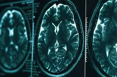
A small-scale study investigated whether the brain's cortical structure could predict how well individuals with major depressive disorder respond to the antidepressant vortioxetine. Researchers used high-resolution MRI and surface-based morphometry to examine the cortex. Findings suggest that specific features of brain regions involved in emotion regulation, planning, and visual processing, as measured before treatment, are associated with improvements in depressive and anxiety symptoms. This exploratory research points towards potential biomarkers for personalized depression treatment, though larger studies are needed to confirm these preliminary results.

Diverse Perspectives on 'Love on the Spectrum': A Reality TV Analysis
This analysis delves into critical discussions surrounding the Netflix reality series 'Love on the Spectrum,' which explores dating among autistic adults. Drawing insights from autistic individuals, journalists, and clinical professionals, the article highlights the show's departure from conventional reality TV drama, its portrayal of kindness and acceptance, and its role in challenging misconceptions about autistic people's romantic lives. It also examines debates over LGBTQ+ representation, family dynamics, the show's realism, and, most notably, the divergent views on dating coaching methods.

Understanding the Perceived Rise in Autism Diagnoses
The increasing rates of autism diagnoses over recent decades have led to public and scientific debates. This article delves into various research studies to differentiate between actual biological increases in autism incidence, improved diagnostic methods, broadened diagnostic criteria, and socio-economic factors influencing reported prevalence. It explores how these elements contribute to the observed rise, examines shifts in demographic profiles of diagnosed individuals, and discusses ongoing debates among experts regarding interpretation of these trends.

The Power of Personal Disclosure: Boosting Likability in Socially Anxious Individuals
New research suggests that sharing personal stories can significantly increase how much others like and want to interact with individuals, especially benefiting those with social anxiety. The study highlights that the listener's personality traits, such as their own social anxiety levels and need for affiliation, also play a crucial role in shaping these positive evaluations, challenging the common perception that only the speaker's behavior dictates social acceptance.