 Breaking News & Live Updates
- Breaking News & Live Updates
- Breaking News & Live Updates
- Breaking News & Live Updates
- Breaking News & Live Updates
- Breaking News & Live Updates
- Breaking News & Live Updates
- Breaking News & Live Updates
- Breaking News & Live Updates
- Breaking News & Live Updates
Short Video Consumption Temporarily Suppresses Cognitive Control Networks, Study Reveals
A recent neuroscientific investigation has shed light on how engaging with preferred short-form video content can lead to a temporary reduction in the activity of brain areas critical for self-regulation and vigilance. This effect, published in the journal NeuroImage, appears to be connected to the concentrations of glutamate, a key neurotransmitter, within the brain.
The Impact of Short-Form Videos on Brain Activity
The study highlights that popular short video platforms, characterized by their rapidly changing, captivating clips, might induce a state where the brain's cognitive control mechanisms are temporarily subdued. While these platforms offer entertainment, researchers are increasingly interested in understanding why some individuals find them difficult to disengage from. One compelling hypothesis suggests that the deeply immersive and rewarding nature of such viewing experiences could diminish the necessity for active self-monitoring and cognitive effort, allowing the brain to enter a more passive state.
This research specifically focused on two crucial brain regions: the dorsal anterior cingulate cortex (dACC) and the dorsolateral prefrontal cortex (dlPFC). The dACC plays a vital role in identifying conflicts, overseeing behavior, and determining when heightened mental exertion is required. Meanwhile, the dlPFC is instrumental in exerting executive control, such as sustaining focus and resisting distractions. Collectively, these areas are fundamental for individuals to manage their actions effectively when attention and self-control are paramount. The findings indicate that when participants watched videos they enjoyed, both the dACC and dlPFC showed a significant decrease in activity compared to their baseline levels. This suggests that during pleasurable short-video consumption, these critical cognitive control networks become temporarily less active, potentially contributing to the difficulty some users experience in limiting their engagement with such content.
Neurochemical Insights and Study Implications
The investigation further explored the role of specific brain chemicals: glutamate, the primary excitatory neurotransmitter that boosts neural activity, and gamma-aminobutyric acid (GABA), the main inhibitory neurotransmitter that helps regulate neural activity. Researchers aimed to determine if resting levels of these chemicals could explain individual variations in how the cognitive control network responds during short-video viewing. The study, led by Tiantian Hong from Zhejiang University, involved 56 young adults, who underwent proton magnetic resonance spectroscopy to measure glutamate and GABA concentrations in the dACC before engaging in a short-video viewing task during functional magnetic resonance imaging (fMRI).
A key discovery was that individuals with elevated resting glutamate levels in the dACC exhibited less suppression of both cognitive control regions during video consumption. Conversely, GABA levels did not show a significant correlation with activity in these areas. The authors concluded that the immersive experience of watching preferred short videos leads to a deactivation of the cognitive control network, and that individual differences in this deactivation are directly linked to glutamate metabolism. While the study found increased connectivity between the dACC and dlPFC during short-video viewing, particularly for liked content, this was interpreted not as a sign of stronger self-control, but rather as a coordinated downregulation of these regions during preferred viewing. The research acknowledges limitations, including the absence of detailed assessment for short-video addiction, the subjective definition of "liked" videos, and a predominantly male sample of young adults, which may limit the broad applicability of these intriguing findings.
Other Articles
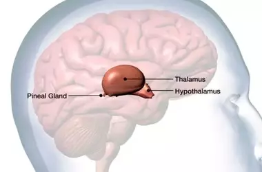
Reduced Pineal Gland Volume Linked to Psychiatric Disorders, Study Finds
A new meta-analysis suggests that individuals with psychiatric conditions like depression, bipolar disorder, and schizophrenia tend to have a smaller pineal gland. This structural difference, which appears to be a neurodevelopmental trait rather than a consequence of illness, may influence melatonin production. However, the study found no direct correlation between pineal gland size and sleep quality, highlighting the complex interplay of factors affecting sleep and mental health.

Antidepressants May Normalize Brain Tissue Changes in Persistent Depression
A new study reveals that antidepressants duloxetine and desvenlafaxine can normalize brain tissue microstructure in individuals with persistent depressive disorder. The normalization appears to be mediated by the reduction in symptom severity, suggesting that initial abnormal tissue changes might be a compensatory response to depression. These findings enhance our understanding of how antidepressants affect the brain and the underlying mechanisms of chronic depression, despite limitations regarding participant attrition and generalizability.

Brain Scans Uncover Links Between Uneven Intelligence and Attention Deficits in Children
A new study reveals that children with ADHD who have a significant disparity between their verbal and nonverbal intelligence scores exhibit more pronounced difficulties with self-regulation and focus. This cognitive gap correlates with reduced blood flow in the frontal brain regions during tasks requiring impulse control, providing insights into the neurological underpinnings of attention deficits. The findings highlight the importance of assessing distinct cognitive abilities in understanding and addressing ADHD in school-aged children.
Deep Brain Stimulation Physically Remodels Brain's Information Superhighway
A recent study suggests that deep brain stimulation (DBS) for severe depression not only changes brain electrical activity but also physically reshapes the brain's white matter. Researchers found that DBS increased the integrity and myelination of nerve fibers in macaques, leading to altered communication across neural networks. These findings imply that long-term therapeutic benefits may come from structural brain remodeling, not just temporary electrical changes.

Brainstem Network Crucial for Attention Identified
A recent study published in Nature Communications has identified an ancient brainstem network essential for filtering distractions and focusing attention. This network, involving inhibitory neurons in the parabigemino-lateral tegmental inhibitory complex (PLTi), helps animals select important spatial information without affecting basic perception or movement. The findings in mice suggest potential new targets for treatments of attention disorders like ADHD, shedding light on a fundamental cognitive skill that transcends species with varying brain complexities.

Understanding ADHD: New Insights from Glutamate Levels in Adolescent Brains
A recent neuroimaging study sheds light on the neurological underpinnings of ADHD in adolescents, focusing on glutamate levels in the medial prefrontal cortex. The study reveals distinct age-related changes in glutamate concentrations among adolescents with persistent ADHD, remitting ADHD, and those without the condition. These findings suggest a potential link between altered brain maturation processes and the clinical course of ADHD, offering new avenues for understanding and potentially treating this neurodevelopmental disorder.